Share this blog post with your friends
Urinalysis is the process of examining urine samples to determine their physical, chemical, and microscopic qualities, which provides essential information on the urinary system’s health. Urine microscopy and culture, on the other hand, go further by detecting bacteria and germs in the urine, which aids in the identification of infections and urinary diseases.
We will look at the goal, procedures, interpretation of findings, and clinical importance of these tests throughout this article, equipping you with the information to better understand and navigate the world of Urinalysis and Urine Microscopy & Culture.
What is Urinalysis and Urine Microscopy & Culture?
Urinalysis is a diagnostic test that examines the physical, chemical, and microscopic properties of urine. It provides valuable insights into the overall health and function of the urinary system. By analysing urine samples, healthcare professionals can detect abnormalities, monitor disease progression, and assess the effectiveness of treatments.
On the other hand, urine microscopy and culture are additional tests that provide more detailed information about the presence of microorganisms in the urine. Microscopic examination involves the use of a microscope to identify and quantify various elements such as red and white blood cells, bacteria, and crystals. It helps in diagnosing urinary tract infections, kidney disorders, and other conditions.

Source: PatientEngage.com
Urine culture, on the other hand, involves incubating a urine sample to identify and determine the sensitivity of any microorganisms present. This test is crucial in diagnosing bacterial infections and selecting appropriate antibiotics for treatment.
Together, urinalysis, urine microscopy, and culture form a comprehensive approach to evaluating urinary health. They play a vital role in diagnosing and managing a wide range of urinary conditions, allowing healthcare professionals to provide targeted and effective treatments.
Overview of Urinalysis
Urinalysis as explained earlier is a valuable screening and diagnostic technique for a variety of urinary and systemic disorders.
The primary purpose of urinalysis is to detect and evaluate various conditions and diseases affecting the kidneys, urinary tract, and other systems. It aids physicians in the diagnosis of urinary tract infections, renal disease, diabetes, and other medical disorders.
During a urinalysis, several components of urine are analyzed. These are some examples:
- Physical features of urine include its colour, look, and odour, which might indicate hydration levels, the presence of blood, or certain metabolic problems.
- Chemical properties: Urine tests are used to determine the amounts of compounds such as glucose, protein, ketones, and electrolytes. Abnormal levels can suggest diabetes, renal failure, or urinary tract problems.
- Microscopic examination: The urine sample is examined under a microscope for the presence of red and white blood cells, bacteria, crystals, epithelial cells, and casts. These results can aid in the diagnosis of urinary tract infections, kidney stones, and other anomalies.
Urinalysis is a non-invasive and very straightforward test that may be used to diagnose and monitor a variety of urinary and systemic disorders. It serves as an important tool in medical practice, aiding in the early detection and management of diseases.
Introduction to Urine Microscopy & Culture
Urine microscopy and culture (MC & S) play a crucial role in identifying bacteria and detecting infections in the urinary system. These tests are essential for diagnosing and treating urinary tract infections (UTIs) and other urinary-related conditions.
Urine microscopy involves examining a urine sample under a microscope to identify bacteria. This allows healthcare professionals to determine if an infection is present and assess its severity. The presence of bacteria in the urine may indicate a UTI caused by various pathogens.
Urine culture is a laboratory technique where a urine sample is incubated to allow bacteria to multiply. This helps identify the specific type of bacteria causing the infection. Urine culture also provides information on the quantity of bacteria, aiding in determining the severity of the infection.

Source: Private GP London
By combining urine microscopy and culture, healthcare professionals can accurately diagnose bacterial infections in the urinary system. This information is crucial for selecting appropriate antibiotic treatments and monitoring their effectiveness. Early identification and targeted treatment of urinary infections can prevent complications and promote recovery.
The Process of Urine Microscopy & Culture
Urine microscopy and culture comprise a set of techniques that are used to correctly identify bacteria and diagnose illnesses in the urinary system:
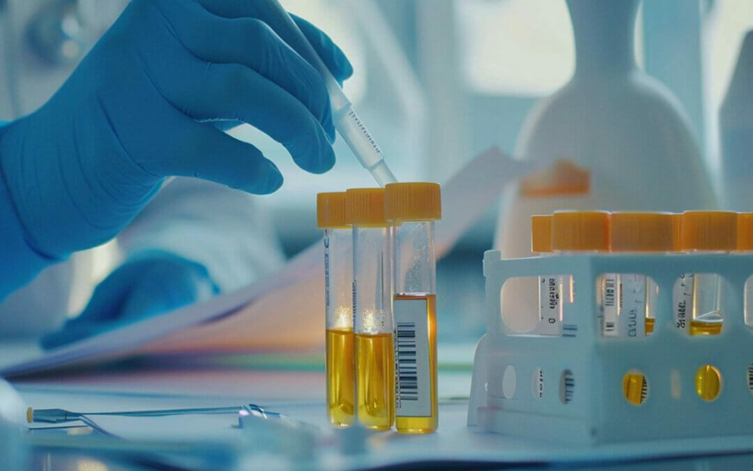
- Collecting the Urine Sample: A clean-catch midstream urine sample is obtained from the patient, while adequate hygiene is maintained to avoid contamination.
- Transporting the Urine Sample to the Laboratory: The urine sample is carried to the laboratory with care, preserving its integrity and avoiding any alterations or contamination.
- Microscopic Examination: A small portion of the urine sample is placed on a glass slide and observed under a microscope. This permits the technician to look for germs, red and white blood cells, epithelial cells, casts, crystals, and other components in the urine.
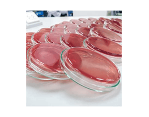
- Culturing the Urine: The remaining urine sample is spread onto agar plates that support bacterial growth. These plates are then incubated at a specific temperature for 24-48 hours.
- Identifying Bacterial Growth: After incubation, the technician examines the agar plates to identify the types of bacteria present. They observe their appearance, colony characteristics, and perform additional tests if needed.
- Antibiotic Sensitivity Testing: If bacteria are found, further tests are conducted to determine their sensitivity to different antibiotics. This helps guide the selection of appropriate treatment.
- The technician collects the findings, including the microorganisms found and their antibiotic sensitivity. These findings are subsequently communicated to the healthcare professional for interpretation and treatment planning.

An Agar plate. Source: Amazon.com
Collecting Urine Samples for Microscopy & Culture
Collecting a clean-catch urine sample is important for accurate urine microscopy and culture results. So, if you were asked to collect one you should follow these steps:
- Wash your hands thoroughly before collecting the sample to prevent contamination.
- Start urinating into the toilet, allowing a small amount to pass first to flush out bacteria.
- Place a sterile urine container under the stream of urine, without touching the inside or lid.
- Collect 30-60 ml of midstream urine, which represents the bladder and reduces contamination.
- Finish urinating in the toilet, excluding the last portion to avoid urethral contaminants.
- Securely close the container lid to prevent leakage.
- Wash your hands again after handling the sample.
Proper handling and storage are crucial:
- Label the container with your name, date, and time.
- If you can’t deliver the sample immediately, refrigerate it at 2-8°C (36-46°F) for up to 24 hours.
- Avoid freezing the sample, as it can affect the results.
By following these guidelines, you ensure reliable urine microscopy and culture results, aiding in the diagnosis and management of urinary tract infections and related conditions.

