Share this blog post with your friends
During urine microscopy and culture (MC & S), a variety of techniques and instruments are utilized to analyze urine samples. These include:
- Microscope: A high-quality light microscope is essential for visualizing and examining urine samples at different magnifications. It enables the identification of cellular components and other microscopic elements.
- Centrifuge: A centrifuge is used to spin urine samples at high speeds, causing solid particles to settle at the bottom of the tube. This process aids in separating the sediment from the liquid portion of the urine, facilitating further analysis.
- Culture plates: Specialized agar plates are employed for urine culture. These plates contain growth media that support the development of microorganisms present in the urine sample. They allow for the identification of bacteria, fungi, or other pathogens responsible for infections.
- Incubator: After inoculating the culture plates with the urine sample, they are placed in an incubator. The controlled temperature and environment within the incubator promote the growth of microorganisms, assisting in their identification.
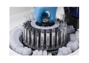
- Automated analyzers: In some modern laboratories, automated urine analyzers are used to perform a range of tests on urine samples. These analyzers can measure various parameters, including pH, specific gravity, protein levels, glucose levels, and more.
 Source: EKF Diagnostics- Automated analyzer (Benchtop)
To achieve reliable analysis, numerous processes are involved in the preparation and inspection of urine samples under the microscope. Here’s a rundown of the procedure:
- Sample Preparation: The collected urine sample is typically mixed well to ensure the components are evenly distributed. A small portion of the well-mixed urine is transferred to a centrifuge tube.
- Centrifugation: The centrifuge tube containing the urine sample is placed in a centrifuge machine and spun at high speed. This process separates the solid components, such as cells, crystals, and bacteria, from the liquid portion of the urine.
- Removal of Supernatant: After centrifugation, the supernatant (the clear liquid above the sediment) is carefully poured or decanted, leaving behind the concentrated sediment at the bottom of the tube.
- Resuspension: A small amount of the concentrated sediment is mixed with a drop of the remaining liquid in the tube. This resuspension ensures that the sediment is evenly distributed for microscopic examination.
- Microscopic Examination: A drop of the resuspended sediment is transferred to a microscope slide, covered with a cover slip, and placed under a microscope. The slide is observed under different magnifications, typically starting at low power (10x) and progressing to higher magnifications (40x or 100x) for detailed examination.
- Observation and Identification: The observer examines the slide for various components, such as red and white blood cells, epithelial cells, casts, crystals, and bacteria. These elements are identified based on their characteristic appearance and morphology.
- Documentation: The findings from the microscopic examination are recorded, which may include the type and quantity of cells, presence of crystals or bacteria, and any other relevant observations. Digital imaging systems can also be used to capture images for documentation and further analysis.
Here are the common Microscopic findings in urine samples after preparation and inspection:
- Red blood cells (RBCs): Their presence may signify urinary tract haemorrhage or kidney-related problems.
- Increased white blood cells (WBCs) indicate inflammation or infection in the urinary tract.
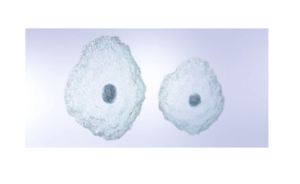
- Epithelial cells: Different kinds of epithelial cells may suggest urinary system injury or abnormalities.

Source: Sysme Europe (Epithelial cells)
- Casts occur when proteins and other chemicals clump together in the kidney tubules, which can indicate a variety of renal disorders.
- Crystals: Certain compounds in urine can form crystals, which may be linked to certain illnesses.
- Bacteria: The presence of bacteria suggests a urinary tract infection or other bacterial illness.
Interpretation of Urine MC & S Results
Interpreting the results of urine microscopy and culture plays a vital role in understanding the health of the urinary system. Let’s explore the various components analysed in urine microscopy and their significance:
Red Blood Cells (RBCs):
The presence of Red Blood Cells (RBCs) in urine, known as hematuria, can have various causes. These include urinary tract infections, kidney stones, bladder or kidney infections, urinary tract trauma, intense exercise, menstrual cycle in women, certain medications, or underlying medical conditions like kidney disease or bladder cancer.
The significance of RBCs in urine microscopy results varies depending on the individual and context. Higher levels may indicate more severe bleeding or an ongoing condition that requires further investigation and management.
RBCs in urine can be associated with different urinary conditions or diseases. They can be a sign of urinary tract infections, kidney disorders, bladder or urinary tract tumours, urinary tract trauma, or blood disorders.
It’s important to note that the presence of RBCs in urine microscopy alone does not provide a definitive diagnosis. Additional tests or consultations with healthcare professionals may be needed to determine the underlying cause and appropriate treatment for the specific urinary condition or disease associated with RBCs in urine.
White Blood Cells (WBCs):
White Blood Cells (WBCs), also known as leukocytes, are of great importance in urine microscopy. They play a vital role in the body’s immune response and can provide valuable insights when observed in urine samples. An increased presence of WBCs in urine, known as leukocyturia, is often indicative of an infection or inflammation in the urinary tract.
When WBCs are elevated in urine microscopy results, it suggests the body’s immune system is actively fighting off an infection. The most common association of increased WBCs in urine is with urinary tract infections (UTIs). UTIs occur when bacteria enter the urinary tract and cause an immune response, leading to an influx of WBCs.
However, it is essential to note that elevated WBCs in urine can also be observed in non-infectious conditions, such as kidney inflammation or autoimmune disorders affecting the urinary system. Therefore, further evaluation and consideration of other clinical factors are necessary to determine the underlying cause and provide appropriate treatment.
If you notice increased WBCs in your urine microscopy results, it is recommended to consult a healthcare professional for proper diagnosis and management.
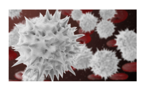
 Red blood cells. Source: istock images
Epithelial Cells:
Epithelial cells are a key component found in urine samples during microscopy. These cells come from the lining of the urinary system, such as the kidneys, bladder, and urethra. They provide insights into the health of these structures.
Different types of epithelial cells, like squamous, transitional, and renal tubular cells, can be observed. Their presence and quantity can indicate specific renal or urinary conditions.
For instance, increased squamous cells may indicate sample contamination, while transitional cells suggest inflammation or infection. Renal tubular cells may signify kidney tubule damage.
Proper interpretation, considering other clinical factors, is essential. Consultation with a healthcare professional ensures accurate diagnosis and appropriate management of epithelial cell-related abnormalities.
Casts:
Casts are structures observed in urine samples under a microscope. They form in the renal tubules and consist of different substances like protein, cellular debris, and minerals.
There are various types of casts, including hyaline, granular, cellular, and waxy casts, each having specific implications. Hyaline casts are commonly found in normal urine, but higher levels may indicate dehydration or kidney damage. Granular casts may suggest kidney tubule injury or chronic kidney disease. Cellular casts, containing red or white blood cells, indicate kidney inflammation or infection. Waxy casts are often seen in advanced kidney disease.
Casts in urine microscopy provide valuable information about kidney-related conditions. However, it’s essential to consider other clinical factors and consult a healthcare professional for accurate interpretation and appropriate management.
Crystals:
Crystals can be detected in urine samples through microscopic examination. They form when certain substances in the urine solidify and clump together.
There are different types of crystals that can appear in urine, such as calcium oxalate, uric acid, struvite, and cystine crystals. Each type of crystal is associated with specific diseases or conditions. For instance, calcium oxalate crystals may indicate kidney stones or hyperoxaluria, while uric acid crystals can be found in conditions like gout or uric acid nephropathy.
Identifying crystals in urine microscopy is clinically significant as it helps diagnose underlying conditions, monitor treatment progress, and guide preventive measures. However, it’s important to note that the presence of crystals alone does not always indicate a pathological condition, as some crystals can be present in healthy individuals too.
To accurately interpret crystal identification, it is essential to consider other clinical information and consult a healthcare professional for proper diagnosis and appropriate management.
Bacteria:
Bacteria can be detected in urine samples through a process called urine culture, where the sample is incubated to allow bacterial growth for identification. This is crucial in diagnosing urinary tract infections (UTIs) and other urinary disorders.
Urine culture helps identify the specific type of bacteria present in the urine, enabling healthcare professionals to determine the most effective antibiotics for treatment. It also involves sensitivity testing to determine which antibiotics will be most successful in combating the particular bacteria causing the infection.
Bacteria play a significant role in UTIs and other urinary disorders. UTIs occur when bacteria enter the urinary tract, leading to inflammation and infection. Kidney infections, bladder infections, and other urinary conditions can also be caused by bacteria. Accurate identification and appropriate treatment of bacterial infections are essential to prevent complications and promote recovery.
It is important to note that the presence of bacteria in urine samples may not always indicate an infection, as contamination during sample collection can occur. Healthcare professionals use urine culture and other clinical information to differentiate between significant bacterial infections and contamination. Consulting a healthcare provider is vital for accurate diagnosis and appropriate management of bacterial-related urinary conditions.
Importance of Urine MC & S in Diagnosing Urinary Tract Infections
Urinary Tract Infections (UTIs)
Urinary tract infections (UTIs) are common bacterial infections that can affect any part of the urinary system. They cause symptoms like frequent urination, pain during urination, and cloudy or bloody urine. Urine microscopy and culture (MC & S) play a vital role in diagnosing UTIs and guiding treatment.
Urine MC & S involves analyzing a urine sample in the laboratory. The sample is cultured to allow bacteria to grow, helping to identify the specific organisms causing the infection. This information is crucial for selecting the most appropriate antibiotics for effective treatment.
By identifying the bacteria and performing sensitivity testing, urine MC & S helps healthcare providers determine which antibiotics will be most effective against the infection. This personalized approach minimizes the risk of antibiotic resistance and increases the chances of successful treatment outcomes.
In a nutshell, urine MC & S is an important tool in the diagnosis and management of UTIs. It helps healthcare providers to identify the microorganisms causing the infection and select the most appropriate medications. This promotes more focused therapy and better patient results.

Source: CDC – A female urinary tract, including the bladder and urethra.
Role of Urine Culture in UTI Diagnosis
Urine culture is a critical component in diagnosing urinary tract infections (UTIs). Its primary purpose is to identify the specific bacteria causing the infection and determine the most suitable antibiotic treatment.
To perform a urine culture, a urine sample is collected and placed in a controlled environment to encourage bacterial growth. The cultured sample is then analyzed to identify the type of bacteria present. Additionally, sensitivity testing is conducted to assess the effectiveness of various antibiotics against the identified bacteria. This information helps healthcare professionals choose the most appropriate antibiotic therapy for the specific infection.
Urine culture aids in the prevention of antibiotic resistance by precisely identifying the microorganisms causing the UTI and selecting the appropriate treatments. This is critical for sustaining antibiotic efficacy and ensuring optimal treatment results.
Briefly, urine culture is critical in UTI diagnosis because it identifies the bacteria causing the illness and guides the selection of appropriate medications. It also helps to prevent antibiotic resistance, ensuring the efficacy of these drugs.
Frequently Asked Questions about Urinalysis and Urine MC & S
Q1: How frequently should urinalysis be performed?
A: The recommended frequency of urinalysis varies based on factors like age, medical history, and specific health conditions. It is typically conducted during routine check-ups, prior to surgeries, or when symptoms related to the urinary system are present. Your healthcare provider will advise you on the appropriate timing for urinalysis based on your individual circumstances.
Q2: Is fasting necessary for urinalysis?
A: Generally, fasting is not required for routine urinalysis. However, specific cases may call for fasting or dietary restrictions, such as when evaluating substances like glucose or ketones in the urine. Your healthcare provider will provide instructions if fasting is necessary for your urinalysis.
Q3: Can medications affect urinalysis results?
A: Yes, certain medications can impact urinalysis results. It is important to inform your healthcare provider about all medications, including over-the-counter drugs and supplements, prior to undergoing urinalysis. This allows them to consider any potential effects of the medications when interpreting the results.
Q4: Can urinalysis detect kidney stones?
A: While urinalysis can provide some indications of the presence of kidney stones, it is not the primary method for diagnosing them. Imaging tests like ultrasound, CT scans, or X-rays are typically used to confirm the existence of kidney stones.
Q5: Can urinalysis detect pregnancy?
A: Urinalysis is not specifically designed to detect pregnancy. Pregnancy tests that detect the hormone human chorionic gonadotropin (hCG) in urine are used for pregnancy confirmation. These tests are separate from routine urinalysis.
Q6: How long does it take to receive urinalysis results?
A: The time required to obtain urinalysis results varies depending on the laboratory and the specific tests being conducted. In some cases, results may be available within a few hours, while in others, it may take a day or longer. Your healthcare provider can inform you about the expected timeframe for receiving your urinalysis results.
Q7: Can urinalysis detect sexually transmitted infections (STIs)?
A: Routine urinalysis is not specifically designed to detect STIs. However, in certain cases, STIs may cause changes in urine that can be identified through urinalysis. If there is a concern about STIs, specific tests targeting those infections are typically recommended.
Q8: Can urinalysis detect drug use?
A: Routine urinalysis is not intended to detect drug use. However, specialized urine drug tests are available to screen for the presence of specific drugs or their metabolites. These tests are distinct from routine urinalysis and are used for purposes such as employment screening or drug therapy monitoring.
Q9: Is a urine sample always necessary for urinalysis?
A: Yes, a urine sample is required for urinalysis. The sample is collected in a sterile container and sent to the laboratory for analysis. Proper collection and handling of the urine sample are vital to ensure accurate and reliable results.
Q10: Can urinalysis be used for monitoring chronic conditions?
A: Yes, urinalysis can be employed to monitor chronic conditions like kidney disease, diabetes, or urinary tract infections. Regular urinalysis helps healthcare providers evaluate disease progression, assess treatment effectiveness, and identify any changes or complications that may arise over time.
How Accurate is Urine MC & S in Detecting Infections?
Urine microscopy and culture (MC & S) are reliable methods for detecting bacterial infections in the urinary system. These tests are highly accurate in identifying the presence of bacteria and determining the specific type causing the infection.
However, some factors can influence the accuracy of the results. Collecting a clean and properly handled urine sample is crucial to obtain reliable results. Contamination or improper storage of the sample can lead to incorrect outcomes.
The timing of the sample collection is also important. Bacterial concentration in urine can vary throughout the day, so a single negative result may not rule out an infection. If symptoms persist, additional testing might be needed.
It’s worth noting that certain antibiotics or antimicrobial agents taken before the test can inhibit bacterial growth in the culture, potentially affecting accuracy. It’s important to inform your healthcare provider about any recent antibiotic use.
Can Urine MC & S Detect All Types of Urinary Tract Infections?
In summary, when performed correctly and under optimal conditions, urine MC & S is a highly accurate diagnostic tool for identifying urinary tract infections. Considering these factors will ensure reliable and accurate results.
Urine microscopy and culture (MC & S) is a helpful test for detecting urinary tract infections (UTIs), but it does have some limitations to consider.
While urine MC & S can identify many types of bacteria causing UTIs, it may not catch every possible pathogen. Certain bacteria may require specific culture conditions or may not show up on standard testing, leading to false-negative results. Moreover, it may not be effective in detecting viral, fungal, or atypical bacterial infections.
It’s important to note that urine MC & S primarily focuses on identifying microorganisms in the urine and may not provide detailed information about the exact location or severity of the infection within the urinary tract.
If UTI symptoms persist despite negative urine MC & S results, further investigations like imaging studies or specialized tests may be necessary to assess the presence of infection.
It’s crucial to communicate openly with your healthcare provider if symptoms continue or if you have concerns about the accuracy of the test. They can guide you on the most appropriate diagnostic approach based on your specific circumstances.
